Skip to content
Guide 015–7 min read

Scoliosis diagnosis: what is measured, how, and why

A scoliosis diagnosis is built from several building blocks. First comes a precise clinical examination and palpation. If indicated, a full-spine radiograph follows, from which the Cobb angle is derived. This page explains which step answers which question.

What is examined in the practice

A structured first clinical examination is performed in underwear. In sequence, the doctor assesses: full-body inspection for pelvic level, shoulder level, and waist asymmetry; trunk rotation measured with a scoliometer during the Adams forward-bend test (ATR value in degrees); spinal mobility; tender points and pain pattern; a neurological baseline status (tendon reflexes, abdominal reflexes, and gross muscle strength); skin findings (café-au-lait spots, hair tufts over the spine, midline skin indentations — all clues to a possible secondary cause); skeletal-maturity signs (menarche in girls; breast development and voice break in boys); and body height measured both sitting and standing. Infobox on the scoliometer: the scoliometer measures trunk rotation in degrees. An ATR value from 5–7° is the threshold at which a radiograph is considered. The value alone is not a diagnosis, but it replaces pure "how it looks" with a reproducible numeric value.

X-ray: once, structured — not ongoing

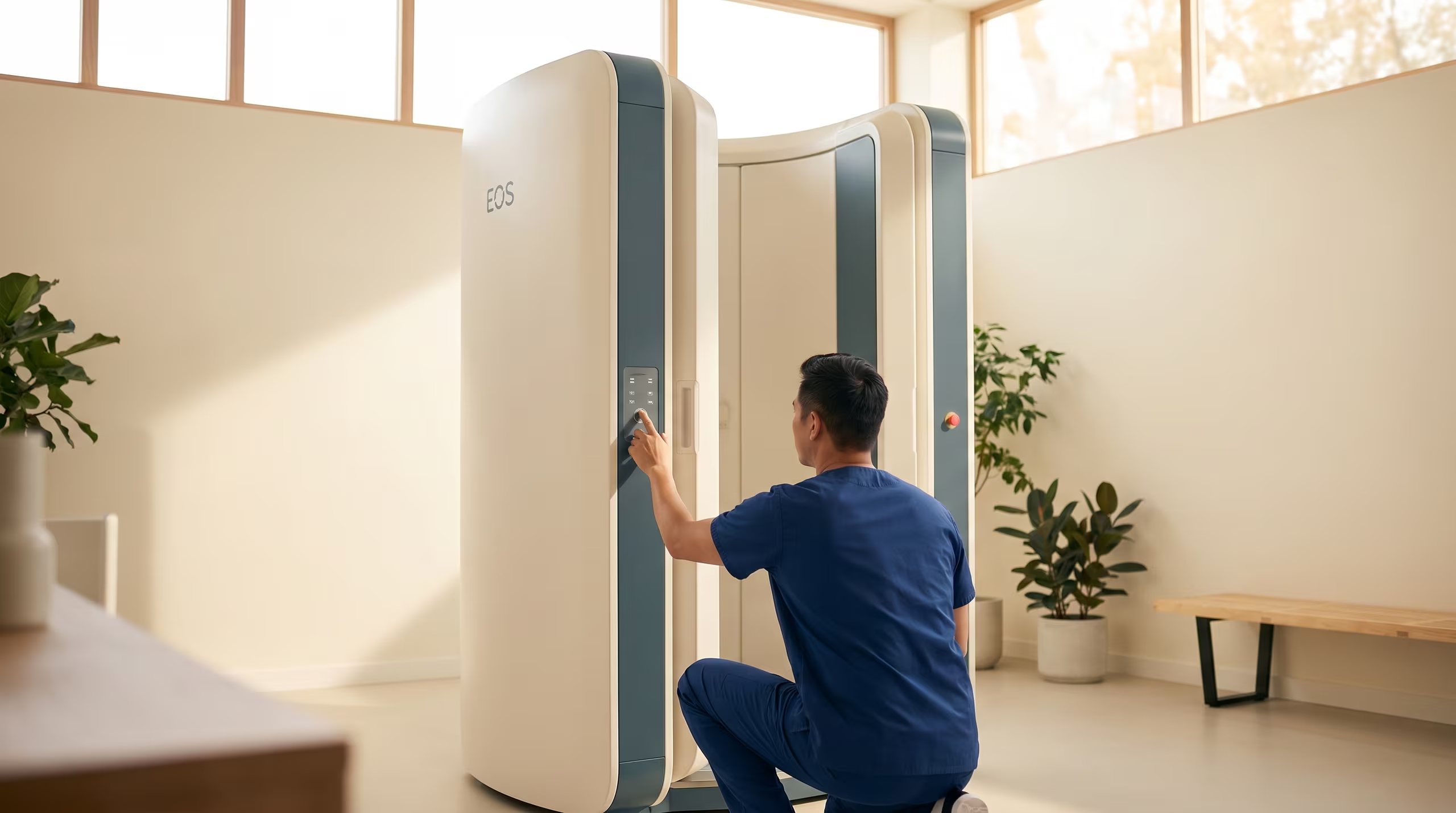
The standard is a full-spine standing radiograph in two planes — posterior-anterior (PA) and lateral. The Cobb angle is measured between the most tilted vertebrae at the top and bottom of the curve. The report notes which side is convex (right-convex or left-convex), which region (thoracic, thoracolumbar, lumbar), and whether a double curve is present. Radiation-dose callout: a full-spine radiograph on modern equipment is low-dose, comparable to a few days of natural background radiation. Follow-up X-rays are deliberately only done when they will change a decision — typically every 6–12 months during growth, more spaced-out otherwise. EOS biplanar systems, where available on site, reduce the dose further. Cobb-threshold infobox (with the four-zone traffic-light framework): under 10°, no disease value and not classified as scoliosis; 10° to under 25° thoracic / under 20° thoracolumbar and lumbar, observation with physiotherapy; 25° to under 40°, indication for brace plus physiotherapy, depending on growth stage; from 40° thoracic and from 40° thoracolumbar/lumbar, surgical consultation, most often surgical indication. Important framing: the thresholds are orientation, not mathematical law. The methodological measurement error is about 3°, and posture-related variation between two X-rays can add roughly another 6°.

EOS low-dose 3D imaging — full-spine scan in seconds

How much growth is still left?

The growth assessment explains the Risser sign (0–5, scored on the pelvic iliac apophysis on the PA radiograph), the Sanders stage (1–8, assessed on a left-hand radiograph for finer resolution in early puberty), and the interval since menarche in girls. Growth potential is the single most important variable for progression risk alongside the current Cobb angle. When the clinical picture requires finer-grained growth assessment — for example in the 25°–40° brace-decision zone — a hand radiograph is added so the Sanders stage can be scored. If growth assessment is not going to change the therapy decision, it is not forced.

MRI — when yes, when no

No routine MRI. Indications are: an atypical curve pattern (for example a left-convex thoracic curve); early-childhood scoliosis (onset under 10 years); neurological findings on exam; rapid progression; suspicious skin findings (café-au-lait spots, hair tufts, midline indentations); strong or nocturnal pain; and planned surgery (preoperative MRI is guideline-recommended). The goal is to rule out a secondary cause — syringomyelia, tethered cord, intramedullary tumors. Where needed, coordination with neurosurgery follows. Outside of these indications, MRI adds cost and anxiety without changing management.

What we hand you after the appointment

After the first visit you leave with a written report — not a verbal "I'll tell you." The report includes: the Cobb angle, curve location (thoracic / thoracolumbar / lumbar), convexity side, rotation findings, skeletal-maturity markers (Risser, Sanders, menarche), key clinical findings, risk assessment, a clear recommendation (observation, physiotherapy, brace, or surgical consultation), and the date of the next check-up. On request we include a short plain-language summary that can be shared with family or another physician.

How often we do check-ups

Reference values: during active growth, clinical check-ups every 3–6 months; X-ray every 6–12 months, or earlier if clinical findings point to relevant progression. After skeletal maturity, longer intervals — usually annually or every two years at stable curve values. A single measurement never drives the decision: what matters is a clear trend across multiple controls under comparable conditions, not a one-off swing within the ~3° methodological error and the ~6° posture-related variability.

For teens: directly to you

If you're here because a doctor mentioned X-rays and measurements — here's the honest version. The X-ray itself is fast: you stand still, it's done in a few seconds, and modern equipment uses a low dose, roughly comparable to a few days of normal background radiation. The Cobb number on the report is a description of the curve, not a label about you. A 22°, a 35°, a 50° — they describe different situations with different plans, not different versions of "how serious a person you are." If the exam shows nothing needs to change, you go home. If something does need attention, you'll get a clear plan and you'll know the timeline. No surprises.

Your Checklist for This Topic

10° Cobb with rotation = scoliosis, but mild curves are often just monitored. The Adams test screens, the X-ray confirms. Cobb measurement has a ~3° error margin. Full-spine standing X-ray in 2 planes is the diagnostic standard. MRI only for pain, neuro symptoms, or pre-surgery.

A scoliosis diagnosis does not mean your child needs surgery. Most mild curves are simply monitored with regular check-ups. The key is understanding the numbers: the Cobb angle tells you the curve size, but growth potential, age, and trend matter just as much. Your job as a parent is to ensure proper monitoring — not to panic about a single measurement.

  1. What is the exact Cobb angle and where is the curve located?
  2. How much growth does my child have remaining?
  3. How often should we schedule follow-up X-rays?
  4. Is this curve likely to progress?
  5. Should we start any treatment now or just observe?

Book a diagnostic appointment at our center — we perform full-spine digital imaging and give you a clear assessment within a single visit.

Book via WhatsApp

Related Guides

Sources & clinical guidelines
  • AWMF-S2kGerman AWMF-S2k Clinical Guideline "Adolescent Idiopathic Scoliosis" (registration no. 151/002), dated 15 March 2023, valid through 14 March 2028. register.awmf.org/de/leitlinien/detail/151-002
  • IQWiGInstitute for Quality and Efficiency in Health Care (IQWiG): information on full-spine X-ray radiation exposure and low-dose protocols.

02 / CONTACT

Book Your Appointment

Message us on your preferred channel. Tell us about your child — we will get back to you with next steps.